ICG Camera & Light
Indocyanine Green (ICG) Cameras, Excitation Light Source and ICG Writing Pen
You can jump to this product in our online store by clicking here

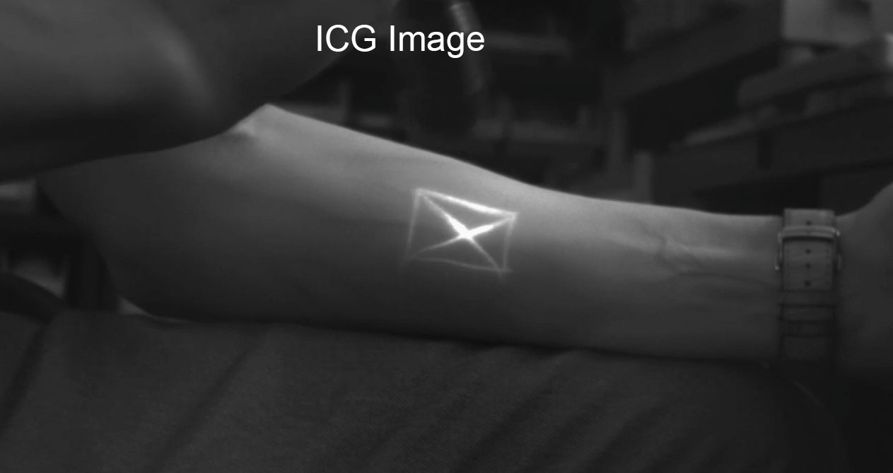
Cost effective equipment for visualizing ICG. Using our ICG handheld light source and one of our ICG cameras, you can quickly, effectively and easily visual ICG. The ICG camera can also be used to visualize vein or vascular structure.
The ICG camera is also compatible with similar dyes that have similar excitation and emissions such as:
- ICG Dye Compatible
- Cypate Dye Compatible.
- IR1 Ink Compatible.
ICG Camera Options
- XNiteICG-USB: USB industrial C-Mount 5 Megapixel camera with 12mm lens, custom ICG filter and software for live video, capturing stills and video and analyzing images. The USB camera is our monochrome C-Mount industrial camera. You can learn more about the camera and its software here.
- XNiteICG-Canon: Canon ELPH 160 20 Megapixel compact camera modified for infrared and with our custom ICG filter
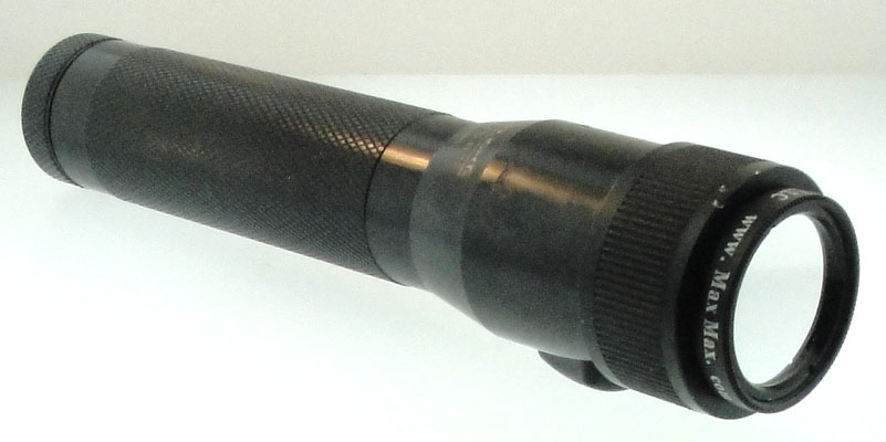
ICG Light Source XNiteICG-Flash
- Handheld
- Portable
- Rechargeable Battery
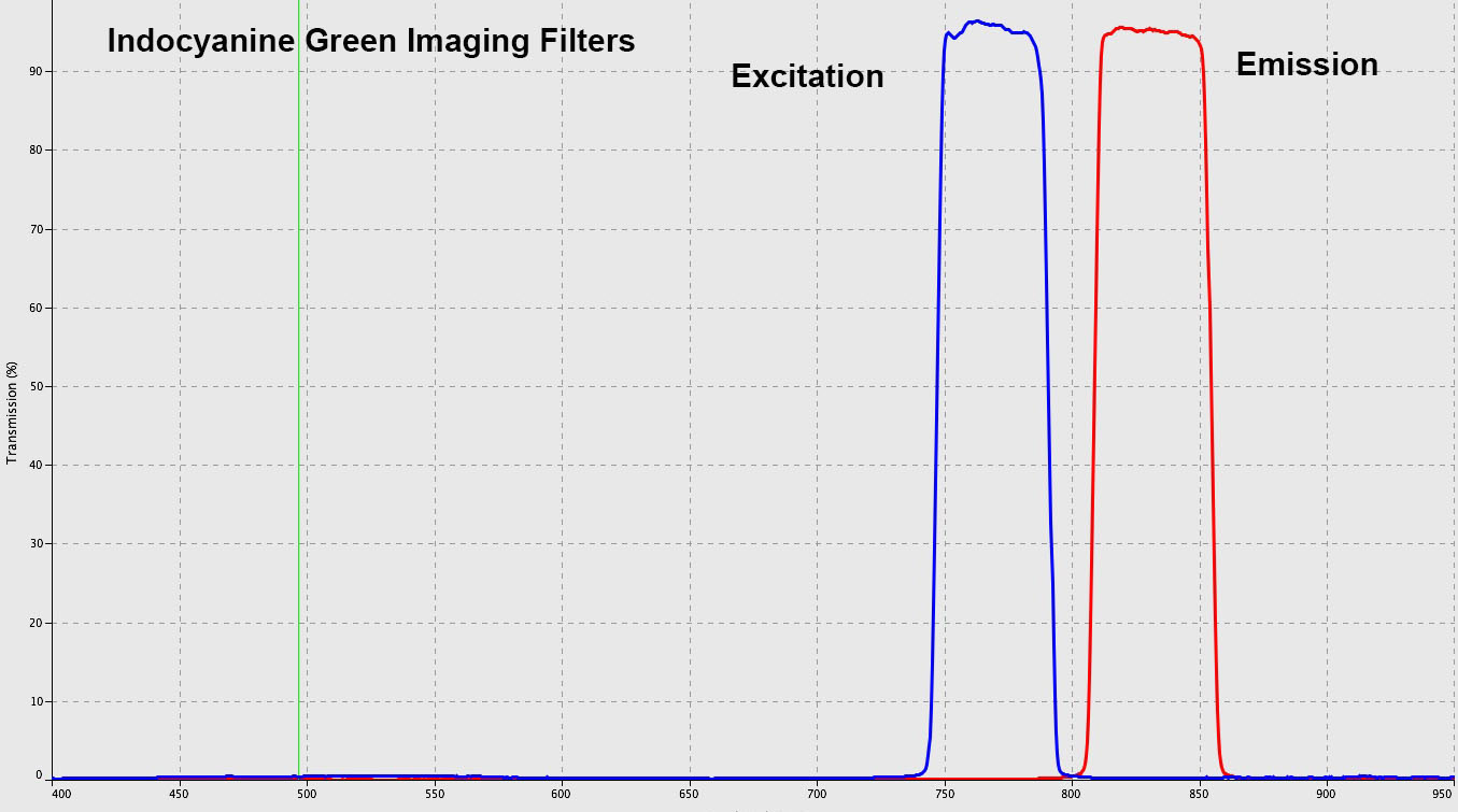
Filter Specifications
ICG Flashlight Excitation Filter
Fluorescence filter typically used for Indocyanine Green imaging for the excitation filter.
90% transmission: 750-789nm
50% transmission: 747-792nm
1% transmission: < 743 and > 796nm
| Center Wavelength CWL (nm) | 769 |
| Bandwidth (nm) | 45 |
| Transmission (%) | 95 |
| Full Width-Half Max FWHM (nm) | 48 |
| Optical Density OD | ≥6.0 |
| OD 6 Blocking Wavelength Range (nm) | 250 - 724 & 808 - 1025 |
| Blocking Wavelength Range (nm) | 250 - 1100 |
| Diameter (mm) | 25.5 |
| Diameter Tolerance (mm) | +0.00/-0.10 |
| Mount Thickness (mm) | 3.5 |
| Thickness Tolerance (mm) | ±0.1 |
| Clear Aperture CA (mm) | 22.0 |
| Transmitted Wavefront, RMS | λ/4 |
| Surface Quality | 60-40 |
| Angle of Incidence (°) | 0 |
| Angle of Incidence Tolerance (°) | ±5.0 |
| Substrate | Fused Silica |
| Typical Applications | ICG-A Excitation |
| Type | Bandpass Filter |
| Coating | Hard Coated |
ICG Camera Filter
Fluorescence filter typically used for Indocyanine Green imaging for the emission filter.
90% transmission: 810-855nm
50% transmission: 812-853nm
1% transmission: < 805nm and > 860nm
| Center Wavelength CWL (nm) | 832 |
| Bandwidth (nm) | 41 |
| Transmission (%) | 93 |
| Full Width-Half Max FWHM (nm) | 41 |
| Optical Density OD | ≥6.0 |
| OD 6 Blocking Wavelength Range (nm) | 550 - 794 & 878 - 1075 |
| Blocking Wavelength Range (nm) | 250 - 1100 |
| Diameter (mm) | 25.5 |
| Diameter Tolerance (mm) | +0.00/-0.10 |
| Mount Thickness (mm) | 3.5 |
| Thickness Tolerance (mm) | ±0.1 |
| Clear Aperture CA (mm) | 22.0 |
| Transmitted Wavefront, RMS | λ/4 |
| Surface Quality | 60-40 |
| Angle of Incidence (°) | 0 |
| Angle of Incidence Tolerance (°) | ±5.0 |
| Substrate | Fused Silica |
| Typical Applications | ICG-A Excitation |
| Type | Bandpass Filter |
| Coating | Hard Coated |
You can jump to this product in our online store by clicking here
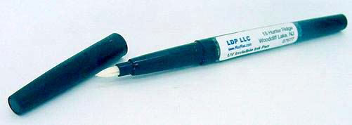
Using our ICG writing pen, you can visualize it with the ICG camera and ICG light

Indocyanine green (ICG) is a cyanine dye used in medical diagnostics. It is used for determining cardiac output, hepatic function, and liver blood flow, and for ophthalmic angiography. It has a peak spectral absorption at about 800 nm. These infrared frequencies penetrate retinal layers, allowing ICG angiography to image deeper patterns of circulation than fluorescein angiography. ICG binds tightly to plasma proteins and becomes confined to the vascular system. ICG has a half-life of 150 to 180 seconds and is removed from circulation exclusively by the liver to bile juice.
ICG is a fluorescent dye which is used in medicine as an indicator substance (e.g. for photometric hepatic function diagnostics and fluorescence angiography) in cardiac, circulatory, hepatic and ophthalmic conditions. It is administered intravenously and, depending on liver performance, is eliminated from the body with a half life of approx. 3–4 minutes. ICG sodium salt is normally available in powder form and can be dissolved in various solvents; 5% (<5% depending on batch) sodium iodide is usually added to ensure better solubility. The sterile lyophilisate of a water-ICG solution is approved many European countries and the United States under the names ICG-Pulsion and IC-Green as a diagnostic for intravenous use.
The absorption and fluorescence spectrum of ICG is in the near infrared region. Both depend largely on the solvent used and the concentration. ICG absorbs mainly between 600 nm and 900 nm and emits fluorescence between 750 nm and 950 nm. The large overlapping of the absorption and fluorescence spectra leads to a marked reabsorption of the fluorescence by ICG itself. The fluorescence spectrum is very wide. Its maximum values are approx. 810 nm in water and approx. 830 nm in blood. For medical applications based on absorption, the maximum absorption at approx. 800 nm (in blood plasma at low concentrations) is important. In combination with fluorescence detection, lasers with a wavelength of around 780 nm are used. At this wavelength, it is still possible to detect the fluorescence of ICG by filtering out scattered light from the excitation beam